Exhibit on Intersections of Medical Science and Art Opens in May 2022

This is a guest post by exhibit curator Sabrina Oliveros 

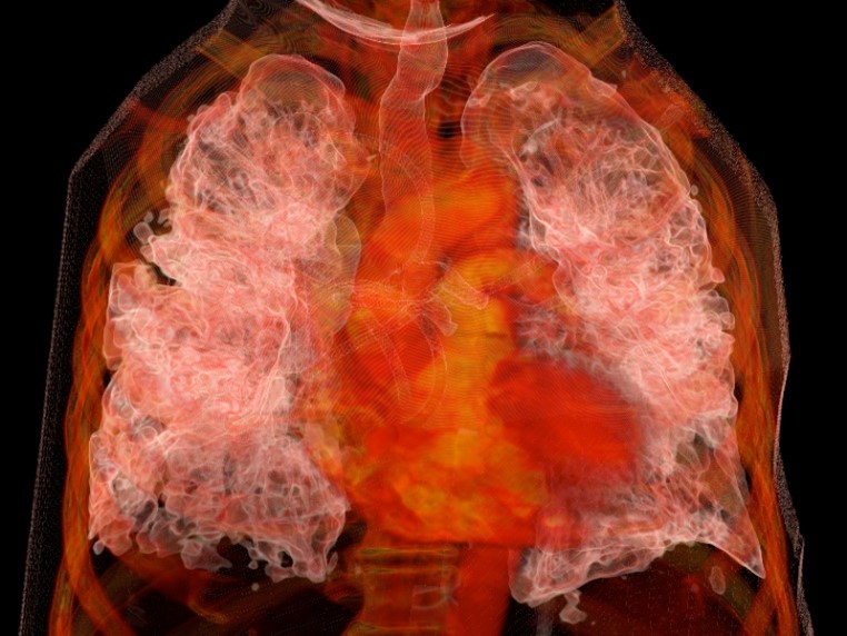
Original engravings from the 17th through the 19th centuries. Facsimiles of masterworks by Andreas Vesalius and Leonardo da Vinci. Images produced through technologies developed at UCSF – including a three-dimensional rendering of a patient’s lungs with COVID-19.

See all these groundbreaking images from the history of medicine, and many more works of science and art, at an exhibit opening in May 2022 at the UCSF Library.

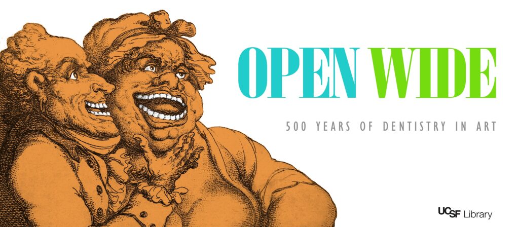
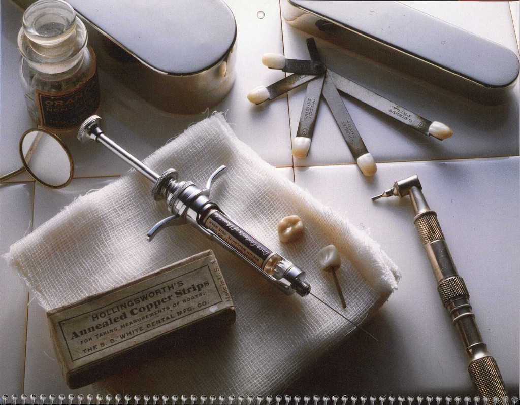
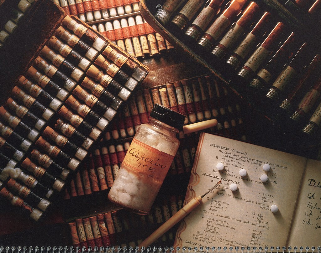
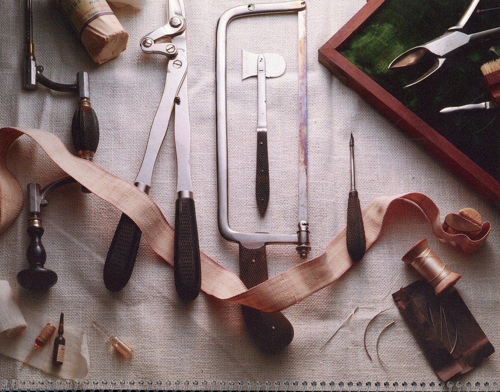
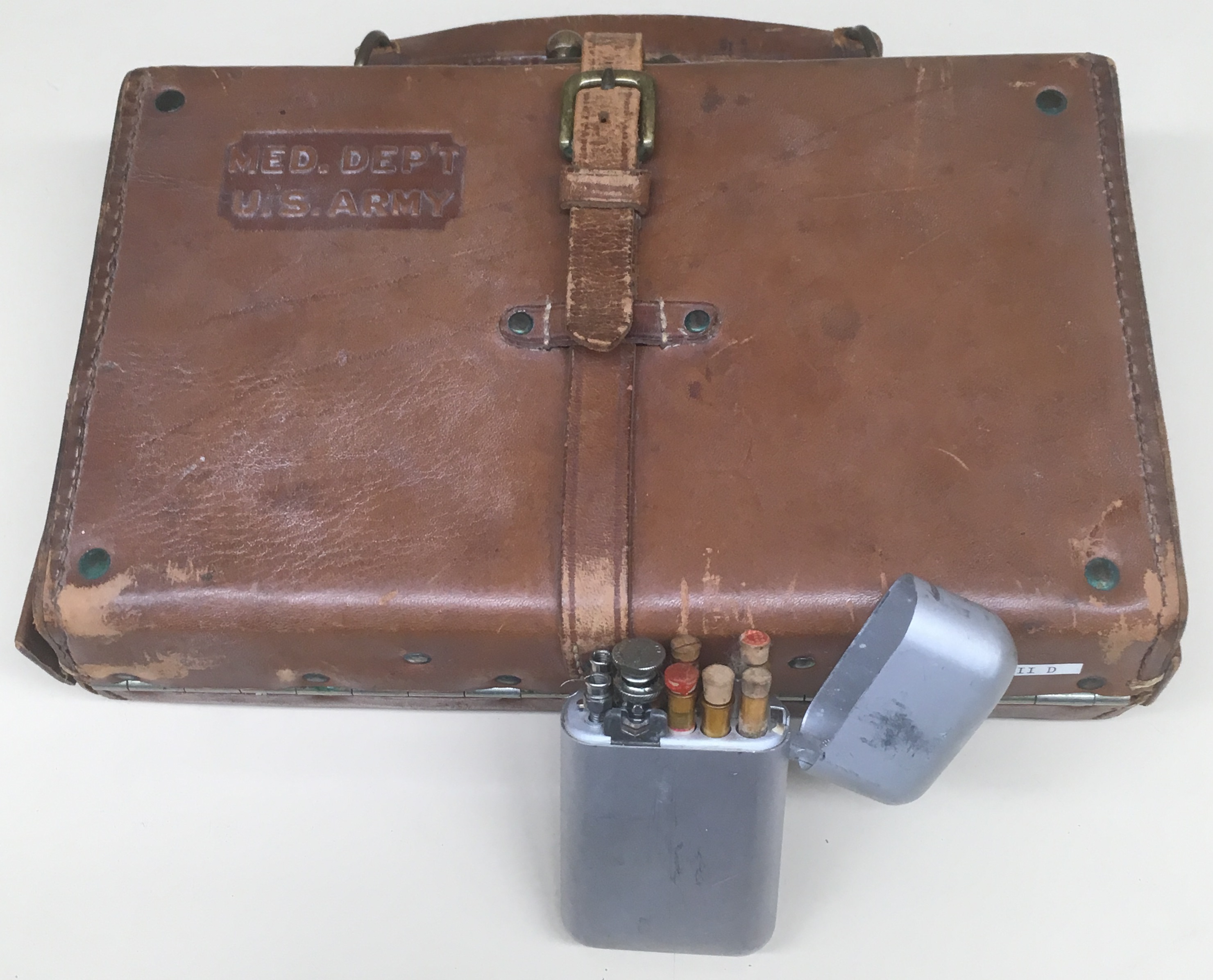
Entitled Seeing the Self Anew: How Art and Science Intersect, this exhibit uses anatomical atlases, medical artifacts, and other materials from UCSF Archives & Special Collections to explore some ways that artists and scientists have informed each other’s work when examining a common subject: the human body.

Frontispiece of De humanis corporis fabrica, libri septem (1543) by Andreas Vesalius.

Collaborations and inspirations

On one level, the relationship between artists and scientists is collaborative. As scientists uncover new knowledge about the body, artists put this information into visual form, recording and disseminating it.

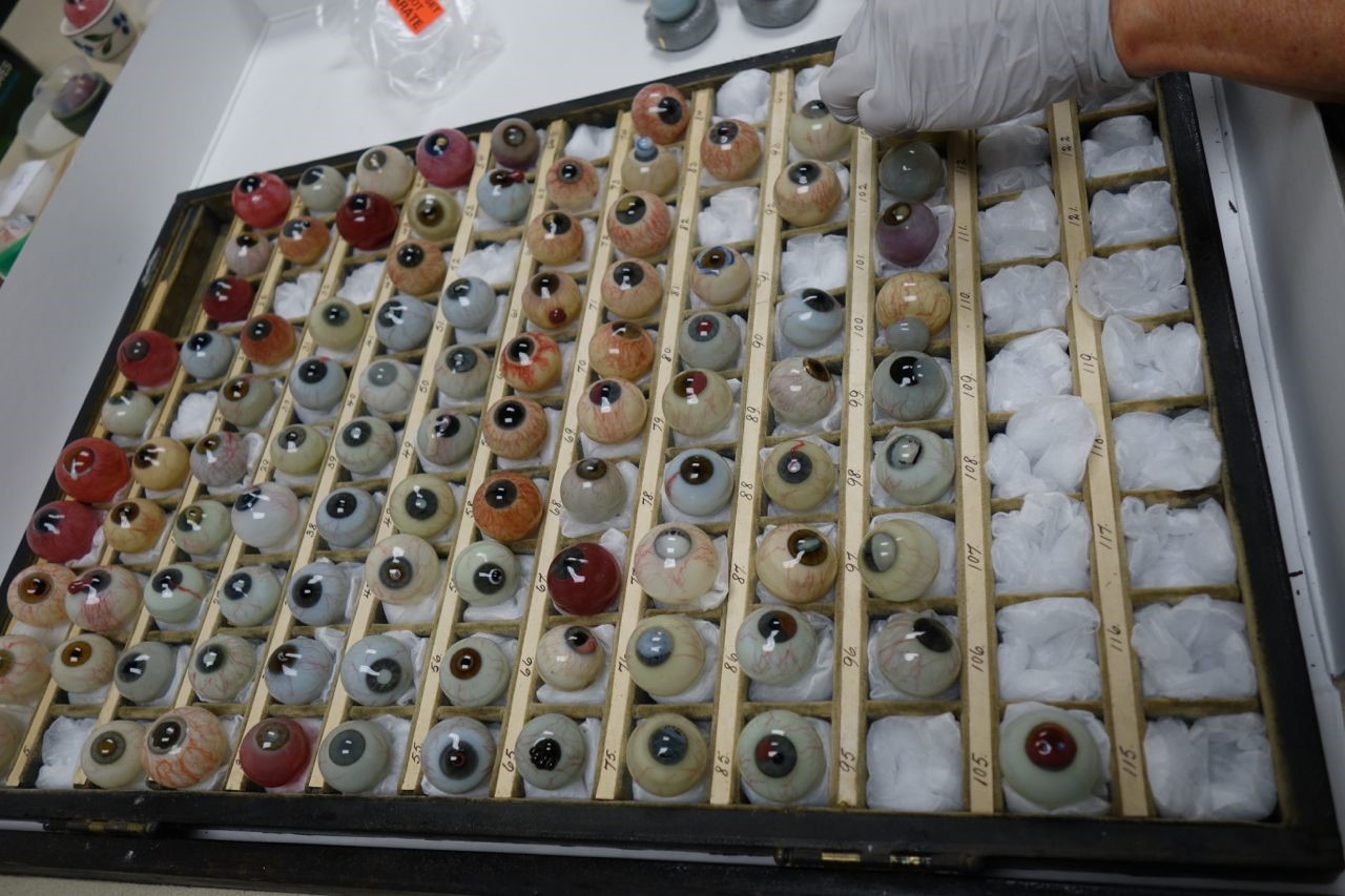
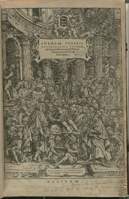
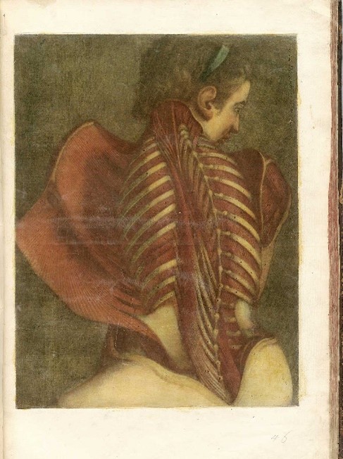
Exhibits in the library’s main lobby feature such collaborations, which, at the times of their publications, counted as the most accurate and attractive anatomical atlases the world had seen. These include books like De humanis corporis fabrica (1543 facsimile) by Andreas Vesalius, which set new directions for the art and science of anatomy; Osteographia (1733) by William Cheselden, which showed the bones in life-like size and detail; and the pioneering manuals on obstetrics by William Hunter (1774) and William Smellie (1793 edition).  

Other displays also show what is created when artists engage with the science of depicting the human body – best exemplified in the works of Albrecht Dürer and Leonard da Vinci.

“The Flayed Angel” from Myologie complètte en couleur et grandeur naturelle (1746) by Jacques Fabian Gautier d’Agoty.

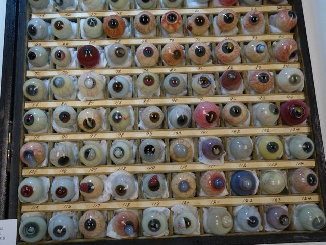
More featured illustrations – like “The Flayed Angel” (1746) by Jacques Fabian Gautier d’Agoty, and fantastical skeletons by Jacques Gamelin (1779) – arguably have less instructional value for medical students. Still, they suggest how anatomical studies inspired artists to produce compelling images all their own.

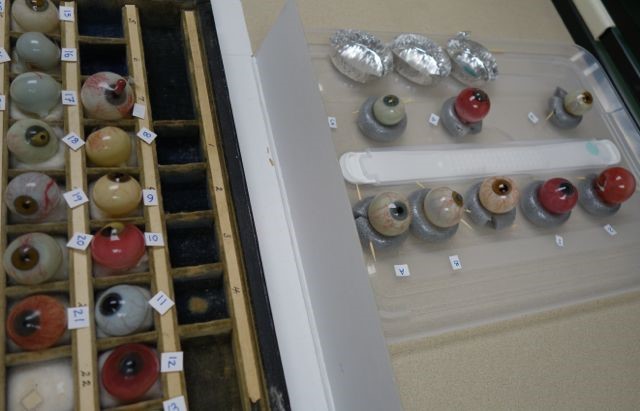
The rare books and artifacts on this floor are supplemented by images from the National Library of Medicine.

Inventions and new dimensions

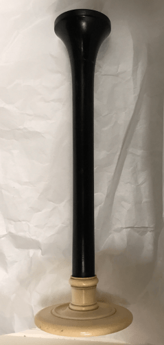
Art and science also intersect in engineering, where drawings guide the design of instruments. From microscopes and stereoscopes to x-rays and MRI technology, these instruments facilitate further studies, procedures, and treatments that produce even newer images of the body.

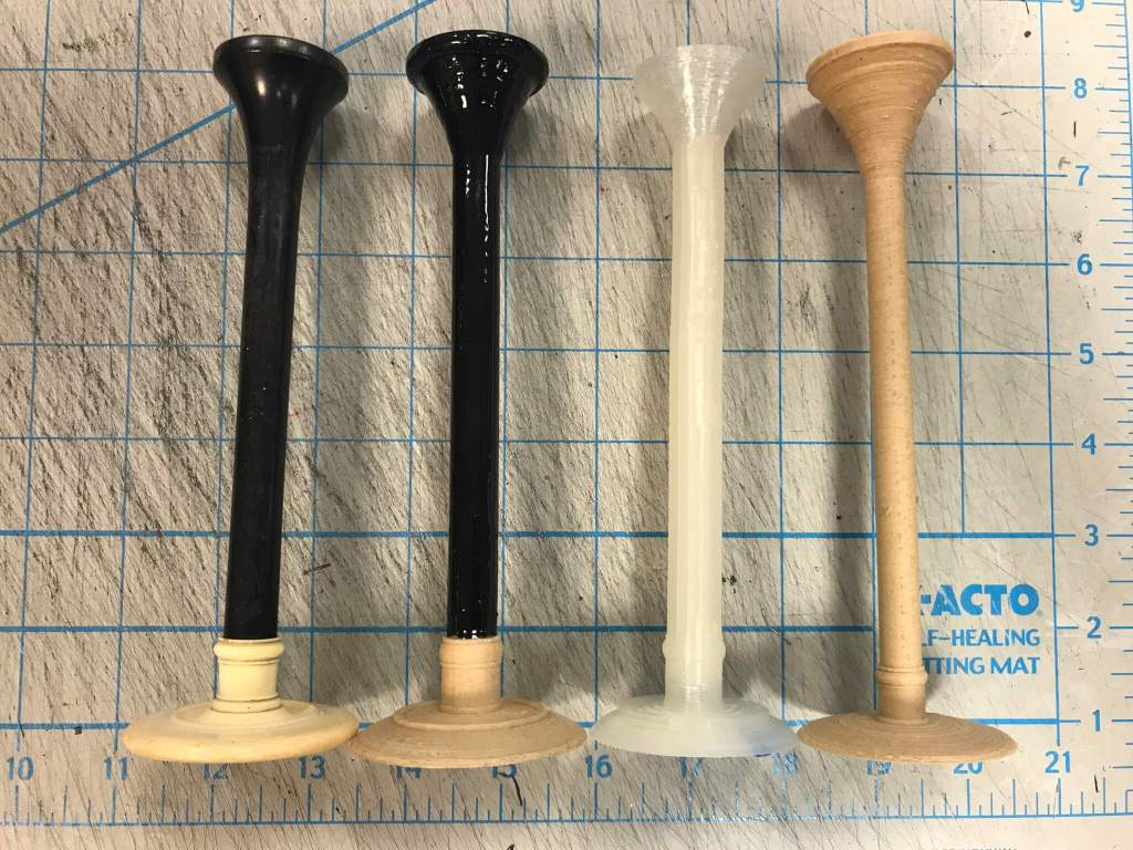
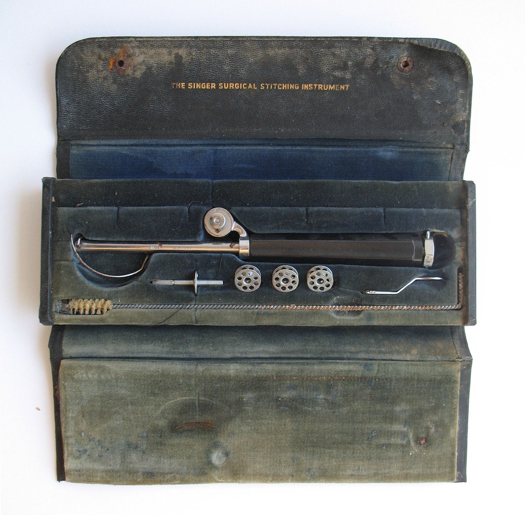
Exhibits on the library’s fifth floor offer a glimpse into such designs. These include conceptual sketches and models envisioned by early 20th-century UCSF professionals like Verne T. Inman, Howard C. Naffziger, and Saxton T. Pope.  

The fifth-floor displays are highlighted by selections from Images, the official publication of the UCSF Department of Radiology and Medical Imaging, dating from 2012 to 2021. With detail and depth that perhaps even the most accomplished early modern artist-anatomists could not have imagined, these illustrations show how far scientists have come – and how much farther they can go – in enabling us to see, and understand, our bodies and our selves anew.

The latest issue of Images magazine renders, in 3D, the lungs of a patient with COVID-19 pneumonia.

Seeing the Self Anew will be on display on two floors (third and fifth) of the UCSF Library at Parnassus through Spring 2023. UCSF Library is currently open to UCSF faculty, staff, and students via ID badge access Monday–Friday from 7:30am–6pm. Changes to in-person library access will be shared through library website as UCSF policies and guidelines are updated in the coming months.

More than Meets the Eye: Restoring the Danz Collection

Guest post by Tracy Power Objects Conservation and Lesley Bone

Funding for this project was generously provided by the Francis I. Proctor Foundation for Research in Ophthalmology at UCSF.

Since 1963, the UCSF Archives & Special Collections holdings have included the historic Danz collection of ocular pathology specimens. The set, one of 13 believed to have been made, was originally intended as a teaching tool for use in medical schools. These blown orbs, some still retaining a long delicate stem, were made in Germany, in the 1880’s, by master glassblower, Amandus Muller. Each glass eyeball depicts, in minute detail, the various diseases and defects that can afflict the eye and is a unique masterpiece of the art of glass making. 

In June 2018 the collection was examined by Tracy Power and Lesley Bone to determine the nature and scope of condition problems that these objects.  Past treatments and current breakages were evaluated, the deterioration of the glass was examined, and current storage conditions were assessed.

While the majority of the glass eyeballs were in stable condition, there were ironically a couple that were themselves suffering from glass disease. This presents with a sticky surface; as a component of the glass leaches out of the surface due to an instability in the glass mix. These surfaces readily attract dust.

Of the previously repaired items, some were in stable condition, but most were in poor condition due to deterioration of the repair materials used and inferior skills of the person or people doing the repairs. One particularly peculiar repair was filled with bright red dental wax.

 The eyeballs were stored in their original compartmented box, with light damaged (faded), velvet-covered cavities for each specimen, and a hinged lid with a glass cover.  The box was still serviceable, but the cavities for the eyeballs had wads of old cotton wool, which was not suitable for the collection since the blown balls retained the thin tubular glass extensions that had been snapped from the rod when the ball was blown. These tended to snag on the cotton.

A treatment plan was agreed upon which would include upgrading the storage container, cleaning all of the glass eyeballs, and repairing the broken glass orbs.

Improved Housing

The eyeballs were removed sequentially for cleaning, and at that time the cavities in the display box were cleaned and new, improved supports were made.  The old cotton wool was replaced with new storage materials that will not be as likely to snag the glass tips.  Small pillows were made of polyester batting in Holytex fabric.  The glass pane in the box was cleaned with detergent and water.  Several discolored areas of paper on the box were toned with conservation stable watercolors and some lifting edges of paper were glued down.

Danz collection of ocular pathology specimens
Old cotton wool was removed and replaced by individually made pillows of archival materials.

Cleaning of the glass eyeballs

Each glass eyeball was carefully cleaned.  A detergent designed specifically for cleaning glass was used for this process.  Handling the eyeballs safely was a major concern and we ended up using foam tubes to make little doughnuts for the glass balls to sit in.  The foam was held in place with toothpicks, so their creation and adjustment was relatively quick. During the cleaning we identified some additional cracks in the glass eyeballs that hadn’t been obvious until they were wet up.  This step was very satisfying as the eyeballs went from dull and cloudy to glistening after cleaning.

Cleaning of the glass eyeballs
Cleaning the glass and the compartments in the box.

Repairing of Glass Eyeballs

Before the eyeballs could be repaired, those with unsightly or failing old repairs had to be undone.  The method varied depending on the types of repair materials previously used.  Several of the repairs had been done with red wax.  The wax remained soft and sticky making it messy and it did not closely resemble glass.  The wax material was removed by gently warming it.  Some of the other old adhesives had failed after becoming brittle.  The brittle material could be brushed from the surfaces, with special care taken to not scratch the glass.  Other old repair materials were removed with solvents.

Repairing the individual eyeballs was the most challenging part of the process, as they are thin and delicate.  Added to that, the high-grade epoxy that was designed for glass conservation can take several days to fully set.  While this can be advantageous, as it allows adjustment of pieces, it also means the fine shards have to be held in place for long periods of time while the resin sets. An advantage of this epoxy is that it is very thin and can be fed by capillary action into cracks.  That property was useful for many of the eyeballs. Also this adhesive has the added advantage of being far superior to commercially available epoxy resins in terms of long-term stability and greater light-stability, therefore it does not yellow like commercially available epoxies. 

Once the eyeballs were repaired, a few had areas where the fragments of the glass were still missing. Glass eyeballs that were incomplete were filled with tinted thermoplastic resin mixtures and details such as veins, were inpainted (inpainting is the process of restoring lost or deteriorated surface decoration or details on an artwork) with commercially ground pigments in acrylic resin.

The glass eyeballs were incredible to work on.   They were beautifully made, if often difficult to look at.  Only one of the eyeballs examined was failing due to unstable glass, or a poor match between the cream under layer and the colored surface glass.  The glass blower had incredible mastery in working with glass in addition to skill in depicting the defects and conditions.  We hope that after this conservation project the glass eyeballs continue to illustrate medical conditions and inspire awe for years to come.

Archives Month

October is American Archives Month, and UCSF Archives and Special Collections is celebrating with a number of events in the coming weeks to showcase our work as custodians of Health Sciences and UCSF History.

Wed October 3: #AskanArchivist Day

Join us and countless other repositories and Archival Institutions on Twitter using the hashtag #Askanarchivist

Pose your burning questions and curiosities about our collections, services and archives work in general.

Follow us on Twitter @ucsf_archives


Wed October 10:

ARCHIVES TALK: Medicine as Mission: Black Women Physicians’ Careers, 1864-1941

Join UCSF Archives & Special Collections as we explore the little-known history of African American women physicians’ careers in medicine from the mid-nineteenth to mid-twentieth centuries. Through an extensive survey of the careers of all known African American women who practiced medicine in this period, a complicated portrait of both accomplishment and constraint emerges. This talk demonstrates that black women physicians succeeded in carrying out their demanding “missions” of attempting to address what we currently term “health disparities” in African American communities. Simultaneously, however, professionalized, scientific medicine in the twentieth century increasingly limited career opportunities available to black women physicians.

Speakers

Meg Vigil-Fowler, PhD is a historian of medicine who studies the intersecting histories of race, gender, and professionalization in the nineteenth and early twentieth centuries. She received her PhD from UCSF’s Department of Anthropology, History, and Social Medicine earlier this year and is currently writing a book on the earliest African American women physicians.

Renee Navarro, MD, PharmD is the Vice Chancellor of Diversity and Outreach, charged with creating and maintaining a diverse university environment where everyone has an opportunity to excel. In her new role, Navarro will collaborate with faculty, staff and students to develop and carry out a strategic plan for diversity and inclusion at the campus – and in recruitment and retention of faculty, students, trainees and staff.

Aimee Medeiros, PhD is an Assistant Professor, History of Health Sciences at UCSF. Medeiros’s work focuses on the reciprocity between diagnoses, preventive care measures, and societal expectations of the body in medicine. Medeiros’s current projects include, Too Young to Die: The History of the Children’s Hospital in the U.S. and Health Sciences Data Laboratory (HSDL), which will complement Big Data efforts by generating historical medical data preserved from non-digital formats.


Saturday October 13: SF Archives Crawl

Join UCSF Archives & Special Collections, California Historical Society, San Francisco History Center, Society of California Pioneers, and Labor Archives and Research Center at San Francisco State University for San Francisco’s second Archives Crawl. The theme for the Archives Crawl is Immigration and Migration to California and we are celebrating in October, which is Archives Month!

Archives Crawl is designed to celebrate archives in the city and encourages guests to explore and engage with institutions that collect archival material. Visit institutions you may not have visited before, pose questions, learn more about what an archive is and what archivists do.

Find the UCSF Archives & Special Collections team at the SFPL Main Branch Library1pm – 5pm

More details visit the San Francisco Archives Crawl site.


Wed October 31

UCSF Archives Halloween Open House: Oddities of the Past

Get in the Halloween spirit and join UCSF Archives and Special Collections on October 31st and view selected pieces from the historical collections in the UCSF Library 5th Floor Reading Room. You will see “medical oddities” of the past including surgical kits, bloodletting tools and more!

Also make sure to drop by the Makers Lab Haunted House anytime from 10am-6pm.


Ongoing exhibit: Open Wide: 500 Years of Dentistry


Pairing Art with Artifact: The Development of Open Wide

This is a guest post by exhibit curator Sabrina Oliveros

Open Wide: 500 Years of Dentistry in Art, which formally opens on September 27 with a reception at the UCSF Library, features a wealth of artworks that depict how perspectives on dentistry, and dentistry itself, have changed over the centuries. The pieces range from satires and caricatures to religious prints and anatomical plates, and they come from artists as different and distinguished as George Cruikshank, Honoré-Victorin Daumier, Francisco de Goya, Utagawa Kuniyoshi, and Marc Chagall.

Remarkable as the art may be, they only comprise half the treasures – and tell part of the stories – in Open Wide.

For this exhibit to find its form, it needed to pair art with artifacts.

Which artifacts could go on display with which artworks? Early project research meant to answer this question.

Which artifacts could go on display with which artworks? Early project research meant to answer this question.

Gateways to learning

Many of the prints in Open Wide had been exhibited from 2003 to 2004 in a show of the same name at the University at Buffalo. When UCSF loaned the artworks from their owner, Dr. Morton G. Rivo, the goal was to expand on the original show using items from Archives & Special Collections. If an artwork illustrated a specific moment in the history of dentistry, the artifacts could elaborate on that moment, helping contextualize what the art showed and turn it into a touchpoint for learning more about the profession.

With some pieces, this task was rather straightforward. The etching Der Zahnzieher (c. 1631-35) by Jan Joris van Vliet (c. 1610 – after 1635), for example, shows a tooth-puller at work; on the wall behind him is a bleeding bowl. Bleeding bowls – which were used to catch drops of a patient’s blood during bloodletting procedures – are among the many historical objects in UCSF’s collections.

Displaying a bowl beside Der Zahnzieher not only added three-dimensionality to the print. It opened an opportunity to discuss why the bowl is in the image and what a tooth-puller used it for (bloodletting was once believed to relieve toothaches). Its presence in the print also suggests that the tooth-puller might have been a barber-surgeon, the kind of tradesman who would have certainly owned such a tool. What is a barber-surgeon and why is this distinction significant in dental practice? Questions and answers can go and on – indicating just how a single artifact can become a gateway into the history of dentistry.

Juxtapositions

The breadth of UCSF’s collections also allowed for other kinds of juxtaposition.

Take one case on the library’s third floor, which contains the print Easing the Toothach (sic). Created long before anything we now use as anesthesia, the image shows a patient who is in such pain that he pulls off his dentist’s wig during treatment. Antique vials of Novocain and an ether gas mask – forerunners of modern local anesthesia – surround the print. In contrast to the bleeding bowl display, the artifacts here expound on the development of dental practice by showing what is absent from the art, not what is visible in it.

Easing the Toothach (sic), by a follower of James Gillray (1757-1815), is the centerpiece of a display on pain management artifacts.

Easing the Toothach (sic), by a follower of James Gillray (1757-1815), is the centerpiece of a display on pain management artifacts.

Another piece on the third floor, the hand-colored engraving Tugging at Eye (High) Tooth (1821), helps showcase a different facet of UCSF’s collections.

 The colorful scene, set in a well-decorated dentist’s office, is by George Cruikshank (1792-1878), one of the most prolific artists during Britain’s golden age of caricature and satire. It shows a dentist furiously at work on a hapless patient, surrounded by his books, dentures, and instruments like teeth-scrapers, a mirror, and a mallet. This piece could have been displayed with similar tools in UCSF’s vaults, again lending three-dimensionality to the office Cruikshank depicts. But there was more to be mined from the print.

Cruikshank lined the dentist’s shelves with titles like Miseries of Human Life, Tales of Terror, and Frankenstein – a tongue-in-cheek suggestion of what the distressed patient is going through. Funny as these were, they raised a few questions: what kinds of books would (or should) have been on a professional dentist’s shelves? And which books shaped the practice so patients would become more comfortable in the chair?

Following this line of thought, the case thus features rare books from the 18th and 19th centuries that advanced knowledge about dentistry. They include the first modern textbook on oral surgery, the first work on orthodontics, and the book that introduced terms like molars and cuspids.

The final third-floor display entitled “The Dentist’s Bookshelf.”

The final third-floor display entitled “The Dentist’s Bookshelf.”

An appropriate addition

Beyond artifacts and rare books, Open Wide also exhibits selections from UCSF’s Japanese woodblock print and School of Dentistry photograph collections. Yet in a university library’s show about dental art and history, perhaps some of the most meaningful materials from the Archives are yearbooks from the school’s early decades.

The Chaff yearbooks displayed on the fifth floor were published from 1897 to 1909 by the junior class of the UC College of Dentistry. They include some truly eye-catching art: one illustration depicts a procedure as an intense sporting match, complete with a referee, spectators, and blow-by-blow commentary; another shows two patients atop a trophy or pedestal, looking like they barely survived a fight. (Its caption? “Patience on a Monument.”)

Such images proved interesting – and unthinkable not to put on exhibit – because they offer historical records of how dental students themselves viewed their profession. More than that, their perspectives surprisingly echo the wry and comical tone of many artworks loaned for Open Wide.

As far as pairing art and artifacts go, there couldn’t have been a more appropriate match than that.

"It is indeed a funny world, But hard truth mingles with the Chaff. It takes some study ere a man May know exactly when to laugh".

A verse from the 1900 volume of Chaff helps explain the spirit behind some yearbook art.


Open Wide Exhibit Opening Reception and Self-Guided Tours

What do a famous French dentist, Snow White, and a Victorian gentleman with a pesky toothache have in common? They are a few of the harassed, horrified, and often hilarious figures you can find in the exhibit Open Wide: 500 Years of Dentistry in Art.

Opening Reception: Thursday, September 27th, 12noon – 1pm, UCSF Library

REGISTER HERE

Join the UCSF Archives and Special Collections for the opening reception and self-guided tours exploring artworks from the collection of Dr. Morton G. Rivo, D.D.S., a former Chief of Periodontics at the UCSF Medical Center at Mount Zion. These selections were first displayed in a 2003 exhibit of the same name at the University of Buffalo. UCSF’s iteration of Open Wide adapts materials from this earlier show and augments the artworks with artifacts, rare books, and UCSF School of Dentistry records from UCSF Archives. Together, they offer a glimpse into how perspectives on dentistry – and dentistry itself – have changed over the years. 

Open Wide will be on display on three floors (first, third, and fifth) of the UCSF Library at Parnassus through August 2019.

12pm Opening Remarks by Dr. Morton G. Rivo, D.D.S., former Chief of Periodontics at the UCSF Medical Center at Mount Zion; Sabrina Oliveros, exhibit curator; and Sara Hughes, MA, EdD. Associate Dean of Education & Student Affairs, School of Dentistry

Guest Curator: Sabrina Oliveros

“Open Wide” exhibit poster

Sabrina Oliveros joined UCSF Archives & Special Collections in April 2018 as the guest curator for Open Wide: 500 Years of Dentistry in Art. Opening this summer, the show will feature selections from the collection of Dr. Morton G. Rivo, D.D.S. that were previously exhibited at the University at Buffalo. Together with artifacts, rare books, and other items in UCSF’s holdings, the artworks will show how perspectives on dentistry – and dentistry itself – have changed through the years.

Sabrina holds a master’s degree in Museum Studies from the University of San Francisco. She co-curated Reformations: Dürer & the New Age of Print at the school’s Thacher Gallery and was the curatorial intern for Company’s Coming: San Francisco Hosts the Panama-Pacific International Exposition at the San Francisco Public Library. She has also been a researcher, scriptwriter, and project assistant for Earprint, an award-winning creator of audio tours, interactives, and immersive sound experiences for museums. Lately, she has been working with the exhibits department at the San Francisco Maritime National Historical Park. Her main jobs are to shore up the Maritime Museum’s research on its WPA-era murals and to develop interpretation for exhibits.

Sabrina Oliveros

Born in New York City and raised in the Philippines, Sabrina has undergraduate degrees in Communication and History from the Ateneo de Manila. She worked for a publishing house and an online marketing firm before venturing into the museum field. Nowadays, when she’s not on exhibit-related projects, she ghostwrites articles for professionals ranging from kitchen remodelers and accountants to dog trainers and, yes – dentists.

3D Printing Artifacts in the Makers Lab

This is an excerpt of a blog post written by Dylan Romero, UCSF Library Makers Lab Manager. Read the full article here.

When the Makers Lab opened in April 2016, we were eager to explore 3D printing applications for UCSF. We soon learned there are countless applications for this technology at a health science institution. Even better, there are some incredible applications for us right here in the UCSF Library, specifically for the Library’s Archives & Special Collections department and the Makers Lab…

Stethoscope from A&SC Health Sciences Artifact Collection

I teamed up with Project Archivist (and Makers Lab volunteer) Kelsi Evans and began to search through the inventory of health science artifacts housed in the Archives. While reviewing the spreadsheet of over 900 items, Kelsi and I continued to find artifact after artifact that we knew had the potential to be recreated in the Makers Lab. Why recreate medical artifacts? Because many of these items are rare, old, and delicate, and must stay behind glass or be closely monitored in the Archives reading room. Why not recreate these artifacts to allow patrons to touch, feel, and interact with the material?

Unlocking the collection was our goal for the proof of concept project. Kelsi and I selected a stethoscope from the 1850’s, made of ebony and bone…

Archives & Special Collections was kind enough to loan the stethoscope to the Makers Lab for the project and I got right to work digitizing the instrument. I began by 3D scanning the stethoscope using the Matter & Form 3D scanner in the Makers Lab.

As you can see in the picture of the original stethoscope, the top potion is black, which unfortunately does not scan well with the 3D scanner. Not a problem, I moved on to modeling the stethoscope using the free, web-based software, Tinkercad. I spent the large majority of my time working in Tinkercad, trying to get the 3D model just right. There is still room for improvement, but the model was good enough for our proof of concept and I was ready to 3D print.

Continue reading the article on UCSF Library’s News page and discover how the printing turned out!

#AskAnArchivist Day – October 4

October 4th is Ask An Archivist Day! We’ll be diligently tending our Twitter account (@ucsf_archives) and responding to questions about our collections, our jobs, or anything else to do with the stewardship of historical material. Tag your questions with #AskAnArchivist to join the conversation.

For example, you could ask:

Have you found any interesting ephemera or artifacts in the AIDS History Project collections? #AskAnArchivist

And we’d say:

Yes, lots. For instance, the papers of epidemiologist Donald Francis include a 12th World AIDS Conference condom and Stop AIDS t-shirt.

12th World AIDS Conference condom, 1998. Donald P. Francis papers, MSS 2015-01.

Stop AIDS t-shirt. Donald P. Francis papers, MSS 2015-01.

#AskAnArchivist Day is part of our October Archives Month programming. Learn more about other upcoming events here.

Health Sciences Artifacts on Calisphere

We’re highlighting one of our recently added digital collections on Calisphere: the Health Sciences Artifact Collection.

The digital collection includes selections from the over 1,000 items in the UCSF Archives Artifact Collection. The items illustrate the development of tools and techniques for medicine, pharmacy, dentistry, and nursing practice.

Of special note are the Advances in Healthcare calendar images. In the late 1980s and early 1990s, UCSF Archives created calendars featuring items from the Artifact Collection. Three of these calendars have been digitized and made available in the Health Sciences Artifact Collection on Calisphere.

Check out all the fascinating items pictured. If you want to take a closer look at any of the artifacts, make an appointment with us to see the real thing in our reading room.

Intern Report: Creating an Exhibit

This is a guest post by Caitlin Toomey, UCSF Archives Intern

Caitlin ToomeyHello, readers! My name is Caitlin Toomey and I was fortunate to be an intern at the UCSF Archives and Special Collections during spring semester. I am currently in the process of receiving my master’s degree in museum studies at USF. Since high school, I have either worked or interned at multiple museums and galleries throughout California, but my time at UCSF stood out as a unique and valuable experience.

While an intern, I was responsible for many different tasks and worked on a number of exciting exhibits. What stood out to me about this internship was the amount of skills I was able to gain and perform throughout the process. For the majority of my internship, I focused on the current exhibit on display in the Library, “DO THE BEST FOR OUR SOLDIERS:” University of California Medical Service in World War I. It was during this time that I completed many different duties.

I began by researching specific subjects, such as the influenza outbreak in 1918 and how troops were entertained on the front, which would be used in the exhibition as stand alone displays. I also wrote the labels with other curators for the exhibit. This was a valuable experience because I mostly have a background in education and collections, so working on more curatorial skills was very helpful. Additionally, collaboratively writing labels can be a challenging but educational experience, and as a result helped me with my writing skills.

WWI exhibit case, “Finding Time to Unwind,” on display in the UCSF Library.

Along with assisting in curation, I was also able to work on exhibit design and collections management for “DO THE BEST FOR OUR SOLDIERS”. I most enjoyed this part of the process because I was able to pick out artifacts for a number of the displays. Looking through the UCSF Archives and Special Collections storage was absolutely fascinating. The collection has so much to explore and discover on the shelves and stacks that I was never at a loss when looking for objects to display. I was also lucky enough to select and help place objects for a number of other special exhibits during my tenure, such as the UCSF Alumni Weekend artifact display of unique health science artifacts and the UCSF Cornerstone demolition series.

WWI-era U.S. Army Medical Department medicine kit used in the exhibit. From the UCSF Archives Artifact Collection, item 218.

Overall, I can look back on my time at the UCSF Archives and Special Collections as a very positive and educational experience. Not many internships give the opportunity to play a large role in exhibitions, as well as learn many different skills that will become valuable for a successful career. I know that I will take with me the many lessons I learned during these past few months. This was a wholly gratifying internship and I will cherish it throughout my career.