Highlights from the Photograph Collection – Radiology Department

Our extensive Historical Photograph Collection includes some really fascinating images. Check out these from the UCSF Radiology Department.

Radon extraction equipment, 1924. Photograph by H.J. Armstrong. Photograph collection, SM, Department of Radiology

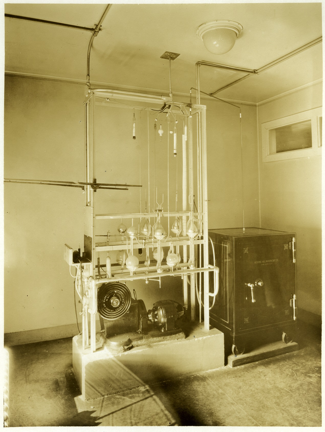
Verso: “Interior of University of California Hospital; Equipment used for the extraction of radon – an element formed by the disintegration of radium,” 1924. Photograph by H.J. Armstrong. Photograph collection, SM, Department of Radiology.

Cobalt machine, 1963; Instrument used to administer radiation therapy, often used for cancer treatment. Photograph by Cal-Pictures. Photograph collection, R, Radiology Therapy.

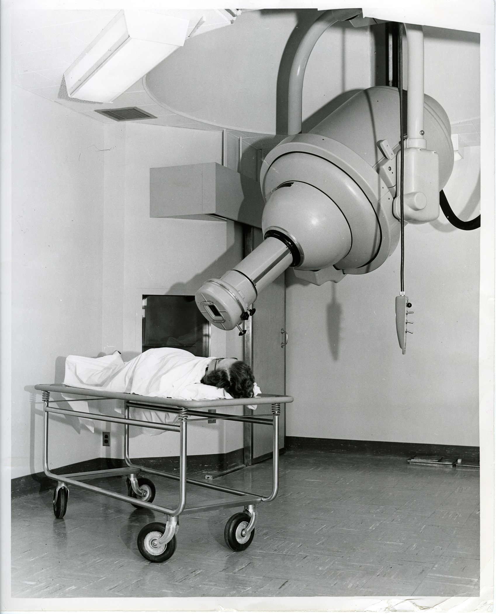
Cobalt machine, 1963. Instrument used to administer radiation therapy, often used for cancer treatment. Photograph by Cal-Pictures. Photograph collection, R, Radiology Therapy.

Students Debbie Modeiros and Nat Rutherford radiologic technology training school practice with doll, 1969. Photograph collection, R, Radiology Technology School.

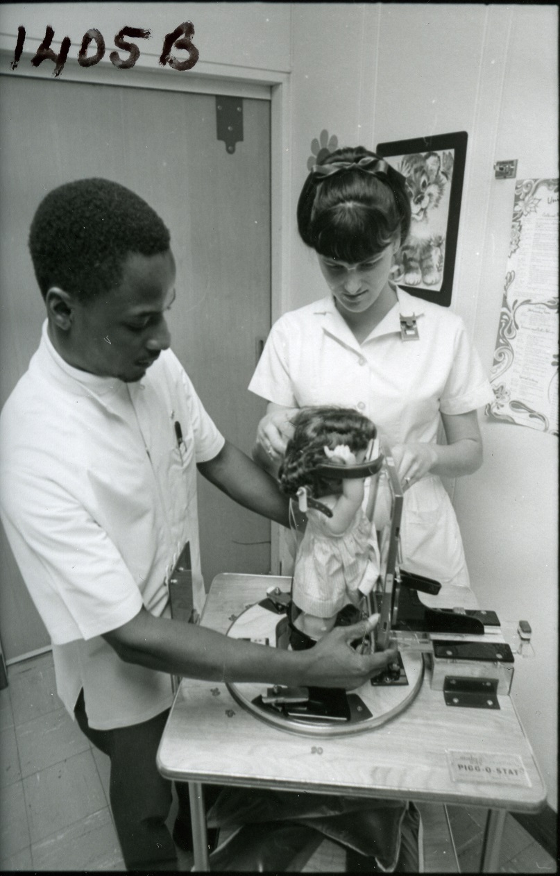
Students Debbie Modeiros and Nat Rutherford, in the Radiologic Technology Training School, practicing with doll, 1969. Photograph collection, R, Radiology Technology School.

EMI Scanner; verso: "The EMI scanner is an example of recent technological advances that reduces patient X-ray exposure while providing more accurate diagnosis than obtainable with older X-ray machines. Here technician Mary McNally and secretary Penny Foster, as patient, demonstrate the giant 2 1/2 ton machine which diagnoses brain disorders." Photograph by Juan Saenz. Photograph collection, R, Radiology CT and EMI scanning.

EMI Scanner, 1975. Verso: “The EMI scanner is an example of recent technological advances that reduces patient X-ray exposure while providing more accurate diagnosis than obtainable with older X-ray machines. Here technician Mary McNally and secretary Penny Foster, as patient, demonstrate the giant 2 1/2 ton machine which diagnoses brain disorders.” Photograph by Juan Saenz. Photograph collection, R, Radiology CT and EMI scanning.

Early MRI Scans

We’re currently processing the Radiologic Imaging Laboratory records, 1968-2000. The collection contains numerous images of magnetic resonance imaging (MRI) scans. The images help document the lab’s achievements in MRI research and illustrate dramatic developments in the technology.

MRI head scan from a patient logbook, 1986, MSS 2002-08. The image is from a Polaroid photograph of a computer screen.

MRI head scan from a patient logbook, 1986. The image is from a Polaroid photograph of a computer screen. MSS 2002-08

MRI scan images come in several formats in the collection. These include marketing prints and slides, transparent film sheets and negatives, and Polaroid photographs. Lab researchers used Polaroid cameras to capture images on computer screens created by in-development software and hardware.

MRI head scan from a patient logbook, 1988, MSS 2002-08. The image is from a Polaroid photograph of a computer screen.

MRI head scan from a patient logbook, 1988. The image is from a Polaroid photograph of a computer screen. MSS 2002-08

Several of the laboratory notebooks in the collection contain Polaroid photographs fastened right to the page, with research notes and data surrounding them.

Laboratory notebook of Lawrence Crooks with scan images, 1983. MSS 2002-08

Laboratory notebook of Lawrence Crooks with scan images, 1983 (subject’s name redacted). MSS 2002-08

As you move chronologically through the collection, you can see the MRI scans becoming clearer and clearer as lab researchers improved the technology. You can also chart changes in the lab’s research subjects. Image subjects transition from phantom objects (containers often filled with baby oil and water) to lab animals and RIL staff and patients.

Prints prepared for a 1985 Diasonics/RIL sales meeting. MSS 2002-08

Prints prepared for a 1985 Diasonics/RIL sales meeting. MSS 2002-08

Though the images present preservation challenges, they contribute greatly to the research value of the collection. Using the scans, you can witness the lab’s growth through different phases of MRI research and development.

Preserving MRI Developments at UCSF

For the next year, I’ll be processing the records of the Radiologic Imaging Laboratory (RIL), 1969-2000. This laboratory pioneered advancements in magnetic resonance imaging (MRI) and helped develop technology that’s now used in hospitals and clinics throughout the world. The collection showcases bioengineering in action and highlights the relationships among industry, research, and education at UCSF.

In process boxes from the RIL records.

In process boxes from the RIL records.

RIL founder and electrical engineer Lawrence E. Crooks gifted the collection to the archives in the early 2000s. It includes over 80 cartons of material ranging from lab notebooks with early scan images to patient records and marketing presentations.

Lawrence Crooks lab notebook, 1979-1983, from the RIL records, MSS 2002-08

Lawrence Crooks lab notebook, 1979-1983. From the RIL records, MSS 2002-08

The material traces the RIL’s growth through different funding agencies and corporate affiliations, including Pfizer, Diasonics, and Toshiba. There are even some personal items, like photographs of lab members celebrating Mardi Gras during a conference in New Orleans!

In process marketing slides from the RIL records. The collection includes various image types, including slides, Polaroids of scan images, negatives, and photographs.

In process marketing slides from the RIL records. The collection includes slides, Polaroids of scan images, negatives, and photographs.

Currently, the collection is cataloged (MSS 2002-08) and has a preliminary inventory, though much of the material lacks intellectual control. My goals are to complete the collection’s processing, create a detailed online finding aid, and digitize a large portion of the material. I will also help curate an exhibit at the UCSF library and a companion online exhibit.

In process tapes from the RIL records. The collection includes various kinds of media, including tapes, floppy disks, and videotapes.

In process tapes from the RIL records. The collection includes tapes, floppy disks, and videotapes.

I’m really excited about the project and hope that it will help users better access the material. The collection is rich in research potential and I can’t wait to see the unique projects it inspires.

Stone’s Synchrotron

This season’s issue of UCSF Magazine, Fall 2013, includes a story inspired by a photograph in our collection and features an audio clip from one of our oral histories.

Robert Stone with Synchrotron, circac 1956

Robert Stone with Synchrotron, circa 1956

This compelling photograph (which, despite appearances, is not a scene from a sci-fi movie) depicts Dr. Robert Stone with the machine he created, the 70MeV electron synchroton. The synchrotron was a type of particle accelerator used to treat cancer patients with radiation from 1956 to 1964. Stone’s work contributed greatly to the safe clinical use of radiation.

The article in UCSF Magazine goes on to elaborate on Stone’s impact here at UCSF and on the wider medical community. The online version of the article also highlights a clip of Stone’s oral history, OH 23, taken in 1964. Be sure to check it out to hear Stone’s story in his own words!

View the issue as an online flipbook, download a pdf, download the iTunes app, or download the Android app. “Robert Stone and the Synchrotron” appears on page 50 of the Fall 2013 issue.

You can read more about Stone’s role in the history of Radiation Oncology at UCSF from 1928-1962 here.