We’re currently processing the Radiologic Imaging Laboratory records, 1968-2000. The collection contains numerous images of magnetic resonance imaging (MRI) scans. The images help document the lab’s achievements in MRI research and illustrate dramatic developments in the technology.
MRI head scan from a patient logbook, 1986. The image is from a Polaroid photograph of a computer screen. MSS 2002-08
MRI scan images come in several formats in the collection. These include marketing prints and slides, transparent film sheets and negatives, and Polaroid photographs. Lab researchers used Polaroid cameras to capture images on computer screens created by in-development software and hardware.

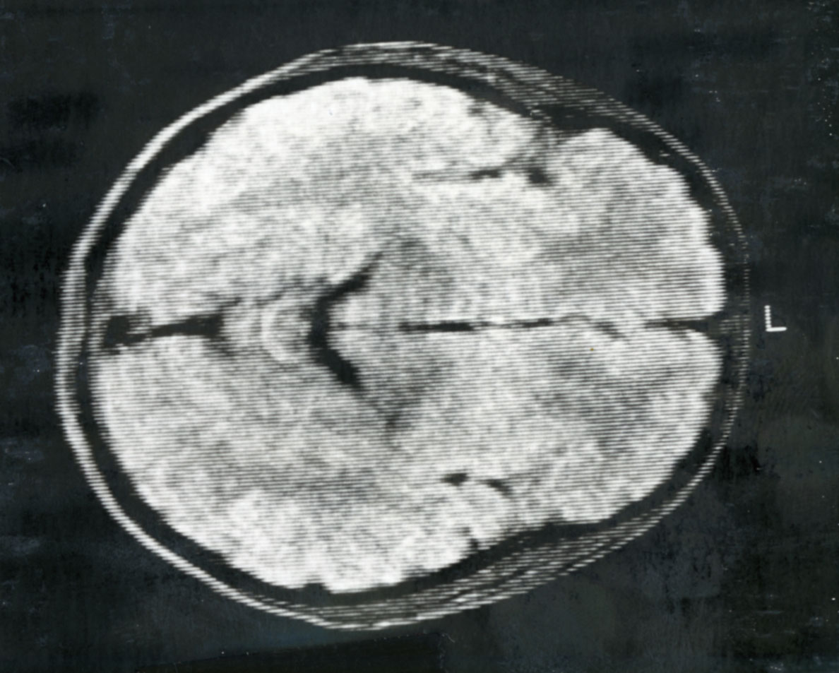
MRI head scan from a patient logbook, 1988. The image is from a Polaroid photograph of a computer screen. MSS 2002-08
Several of the laboratory notebooks in the collection contain Polaroid photographs fastened right to the page, with research notes and data surrounding them.
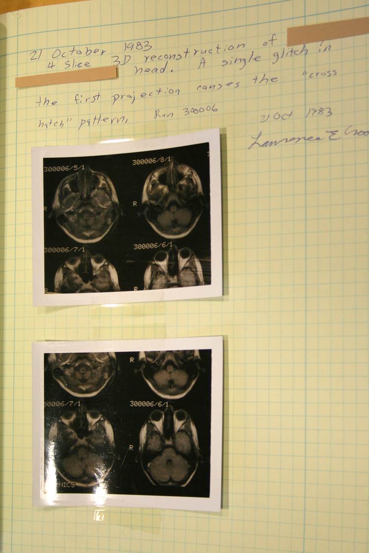
Laboratory notebook of Lawrence Crooks with scan images, 1983 (subject’s name redacted). MSS 2002-08
As you move chronologically through the collection, you can see the MRI scans becoming clearer and clearer as lab researchers improved the technology. You can also chart changes in the lab’s research subjects. Image subjects transition from phantom objects (containers often filled with baby oil and water) to lab animals and RIL staff and patients.
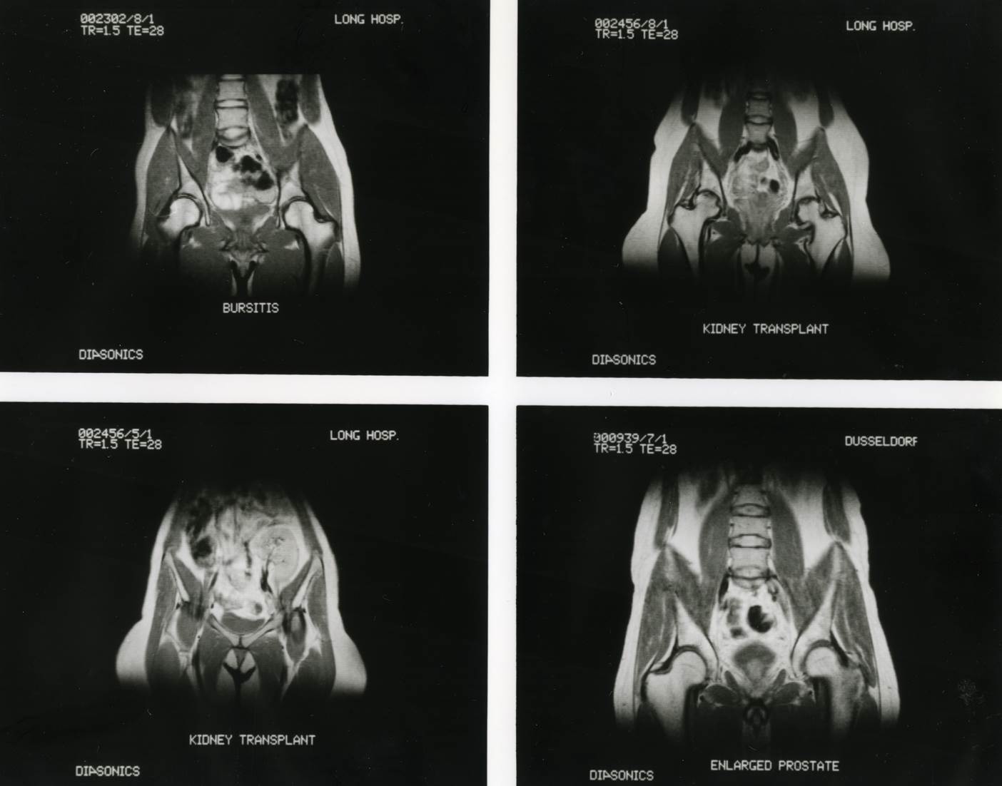
Prints prepared for a 1985 Diasonics/RIL sales meeting. MSS 2002-08
Though the images present preservation challenges, they contribute greatly to the research value of the collection. Using the scans, you can witness the lab’s growth through different phases of MRI research and development.